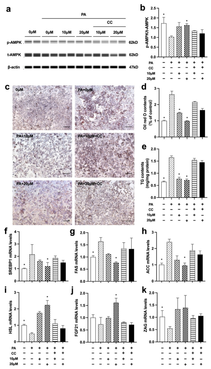
Figure 6.
Nuci prevented PA-induced cellular steatosis by activating AMPK phosphorylation in HepG2 hepatocytes. HepG2 hepatocytes were cultured and treated according to the scheme described in Method 2.9. The protein levels of p-AMPK, t-AMPK, and β-actin were analyzed by Simple western (a), the expression of p-AMPK was normalized against t-AMPK (b). Oil red O staining was conducted and photographed at 100× magnification (c) and Oil red O dye was extracted and detected (d). Intracellular TG contents I were determined by a commercial kit following the manufacturer’s instructions. The mRNA expressions of SREBP1 (f), FAS (g), ACC (h), HSL (i), ZAG (j), and FGF21 (k) were determined by RT-qPCR. Data were presented with mean ± SE from three independent experiments, * p < 0.05 vs. the PA group (PA + 0 μM).

