Figure 7.
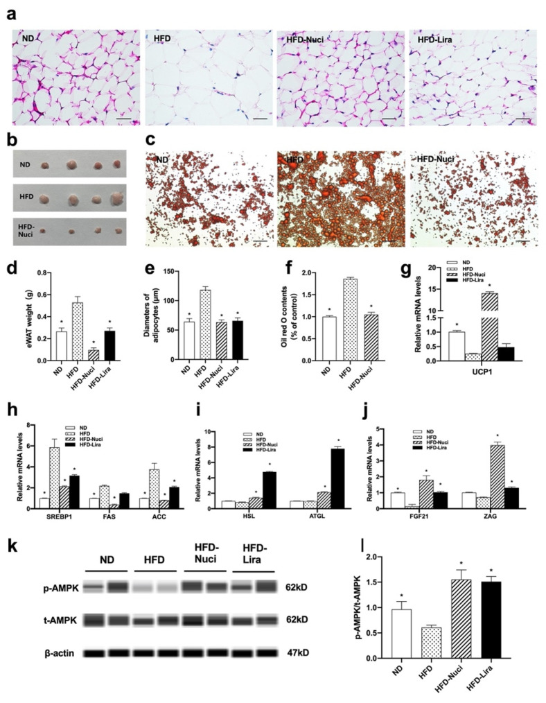
Nuci decreased the weight of eWAT by activating AMPK phosphorylation in HFD-fed mice. Seven-weeks male C57BL/6J mice were fed with ND, HFD, HFD supplemented with Nuci and HFD with subcutaneous injection of Lira at 200 μg/kg/day for 12 weeks. H&E-staining images of representative sections of mice eWAT in the ND, HFD, HFD-Nuci, and HFD-Lira group, photographs were taken at 400× magnification (a). Diameters of adipocytes were measured from six random fields using Image J software (e). The weight of eWAT was measured (d). Photographs of isolated mice eWATs in the ND, HFD, HFD-Nuci group (b). Representative images of the Oil red O stained mice primary mature adipocytes, photographs were taken at 100× magnification (c) and Oil red O dye was extracted and detected (f). The mRNA expressions of UCP1 (g) lipogenesis-related genes SREBP1, FAS, and ACC (h), lipolysis-related genes HSL and ATGL (i), adipokines FGF21 and ZAG (j) were determined by RT-qPCR. The protein levels of p-AMPK, t-AMPK, and β-actin were analyzed by Simple western (k), the expression of p-AMPK was normalized against t-AMPK (l). Data were presented as mean ± SE, n = 12 in each group. * p < 0.05 vs. the HFD group.

