Figure 1.
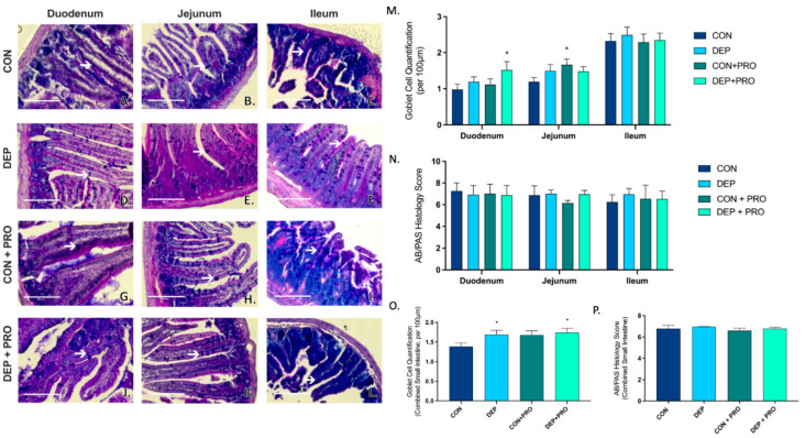
Exposure to inhaled diesel exhaust particles promotes goblet cell formation in the small intestine of C57Bl/6 male mice. Representative images of AB/PAS staining in the three regions of the small intestine of C57Bl/6 male mice on a high-fat diet exposed to saline (CON; A–C), diesel exhaust particles (DEP-35 µg PM; D–F), saline and probiotics (CON + PRO, 0.3 g/d of Ecologic® Barrier probiotics; G–I), or diesel exhaust particles and probiotics (DEP + PRO, 0.3 g/d of Ecologic® Barrier probiotics; J–L) twice a week for 4 w. Panels show representative images within the duodenum (A,D,G,J), jejunum (B,E,H,K), and ileum (C,F,I,L). Graph (M) shows the quantification of goblet cells per 100 µm of intestinal villi by region and (O) shows the global (cumulative quantification of three portions) quantification of goblet cells per 100 µm of intestinal villi in the small intestine. White arrow indicates goblet cell. Graph (N) shows the regional histological mucus score, and (P) shows the global (cumulative) histological mucus score. 40x magnification, scale bar = 100 µm. Data are depicted as ± SEM with * p < 0.05 compared to CON.

