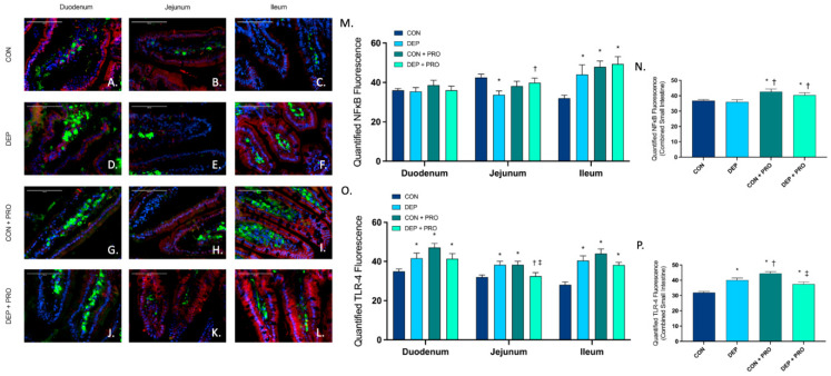
Figure 8.
Exposure to inhaled diesel exhaust particles stimulates TLR-4 expression but not NF-κB in the small intestine of C57Bl/6 male mice. Representative images of NF-κB and TLR4 expression in the three regions of the small intestine of C57Bl/6 male mice on a high-fat diet exposed to saline (CON; A–C), diesel exhaust particles (DEP-35 µg PM; D–F), saline and probiotics (CON + PRO, 0.3 g/d of Ecologic® Barrier probiotics; G–I), or diesel exhaust particles and probiotics (DEP + PRO, 0.3 g/d of Ecologic® Barrier probiotics; J–L) twice a week for 4 w. Panels show merged images within the duodenum (A,D,G,J), jejunum (B,E,H,K), and ileum (C,F,I,L). Red fluorescence indicates NF-κB expression, green fluorescence indicates TLR-4 expression, yellow indicates co-localization of NF-κB and TLR-4, and blue fluorescence indicates nuclear staining (Hoechst). Graph (M) shows the histological analysis of NF-κB by region, (N) shows the global (cumulative expression of three portions) analysis of NF-κB expression in the small intestine, (O) shows the histological analysis of TLR-4 by region, (P) shows the global (cumulative expression of three portions) analysis of TLR-4 expression in the small intestine 40x magnification, scale bar = 100 µm. Data are depicted as ± SEM with * p < 0.05 compared to CON, † p < 0.05 compared to DEP, and ‡ p < 0.05 compared to CON + PRO.

