Figure 1.
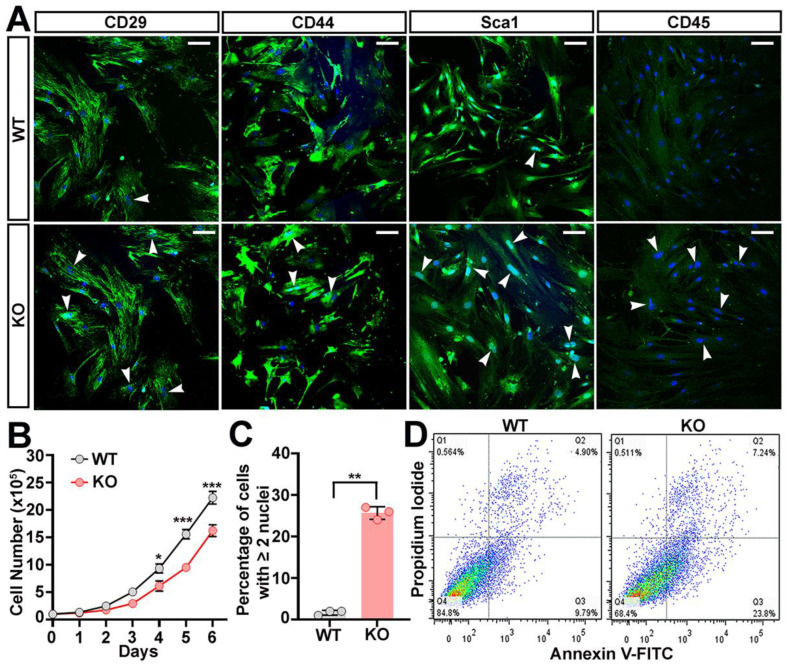
Compromised self-renewal of ASCs cultured from trappc9-null mice. ASCs were isolated from WT and trappc9-deficient mice and cultured as in Methods. Both WT and KO ASCs at passage-3 were subjected to analyses. (A) Confocal images showed that ASCs isolated from WT and trappc9-null (KO) mice expressed mesenchymal stem cell markers and were devoid of hematopoietic CD45. Arrowheads point to cells containing two or more nuclei. Scale bars: 100 μm. (B) Comparison of the growth rate between WT and trappc9-deficient (KO) ASCs. ASCs were cultured at a density of 1 × 10⁵ in T25 flasks and counted every 24 h for 6 days. Cell counting was performed with a hemocytometer. (C) Quantification of cells with two or more nuclei revealed that trappc9-null (KO) ASCs were defective in cell division. Images captured from 3 randomly chosen visual fields from cells for each genotype were used for cell counting. (D) Apoptosis of cells in WT and trappc9-deficient (KO) ASC cultures was examined by flow cytometry-based analysis of phosphatidylserine exposure (Annexin V-FITC staining) and plasma membrane integrity (propidium iodide staining). Shown are dot plot graphs from one of three individual analyses. Similar observations were achieved in other two experiments. Data in (B,C) are Mean ± SD. Each symbol represents one experiment. Statistical significance was determined by unpaired two-tailed Student’s t-test: * p < 0.05; ** p < 0.01; *** p < 0.001.

