Figure 5.
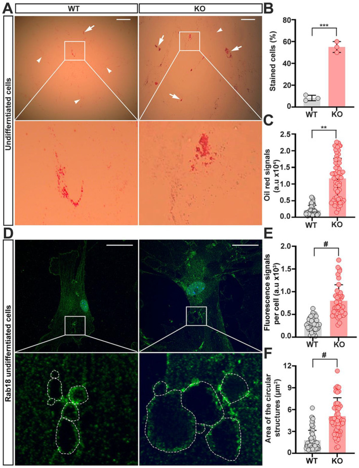
Enlargement of lipid droplets occurs in un-induced trappc9-deficient ASCs. WT and trappc9-deficient ASCs at passage-4 were seeded in 6-well plates with or without glass coverslips and cultured overnight. Cells in wells without glass coverslips were stained with oil red, whereas cells on glass coverslips were processed for immuno-labeling rab18. (A) Representative images of oil red stained cells. Enlarged boxed regions beneath the corresponding image showed clusters of enlarged lipid droplets in un-induced trappc9-null (KO) ASCs. Arrows identified cells having clustered large lipid droplets, whereas arrowheads indicated cells lacking such clustered lipid droplets. Scale bars: 100 μm. (B) Cell counting revealed more trappc9-defidient (KO) ASCs displaying clustered lipid droplets than WT ASCs. For each of 3 independent experiments, 100 cells were counted from 3 images taken from randomly chosen and non-overlapped visual fields. (C) Densitometry of oil red signals suggested greater accumulation of lipids in trappc9-null (KO) ASCs than in WT ASCs. (D) Representative confocal images of cells labeled with antibodies for rab18. Dashed contours in enlarged boxed regions beneath the corresponding image indicated circular structures formed by rab18-positive signals. Scale bars: 10 μm. (E) Densitometry of rab18-immunoreactive signals revealed upregulation of rab18 in trappc9-deficient (KO) ASCs. (F) Measurement of the size of circular structures in cells as highlighted in images (D), showed enlargement of rab18-positive structures in trappc9-deficient (KO) ASCs. The size of rab18 decorated circular structures from 15 cells for each genotype was measured. Data are Mean ± SD. Each symbol represents one experiment in (B), one cell in (C,E), and one circular structure in (F). Unpaired two-tailed Student’s t-test: ** p < 0.01; *** p < 0.001; # p < 0.0001.

