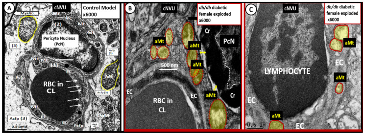
Figure 10.
The mural pericyte (Pc) and endothelial cell (EC) of the capillary neurovascular unit (NVU) develop remodeled aberrant mitochondrial (aMt) in the 20-week-old female diabetic db/db models. Panel A demonstrates the normal electron-dense mitochondria (Mt) in the Pc and EC (outlined with white lines) in the cytoplasm. Additionally, note the arrows pointing to the tight and adherens junctions and normal electron-dense Mt in the astrocyte foot processes (outlined in yellow). Panel B depicts the aMt (pseudo-colored yellow outlined in red) in the Pc cytoplasm as compared to the normal Mt in panel A. Panel C depicts the aMt in the EC (pseudo-colored yellow outlined in red) as compared to the normal Mt in panel A. Note the excessive chromatin (Cr) condensation in the Pc nucleus. Panels B and C are exploded cropped images and these images were observed during the studies in references [32,33,34,35,36] provided by CC 4.0. Original magnification ×6000 exploded with intact scale bars of 500 nm (panel B) and 0.5 μm (panels A and C). Acfp = astrocyte foot process; CL = capillary lumen; Pcfp = pericyte foot processes; PcN = pericyte nucleus; RBC = red blood cell(s).

