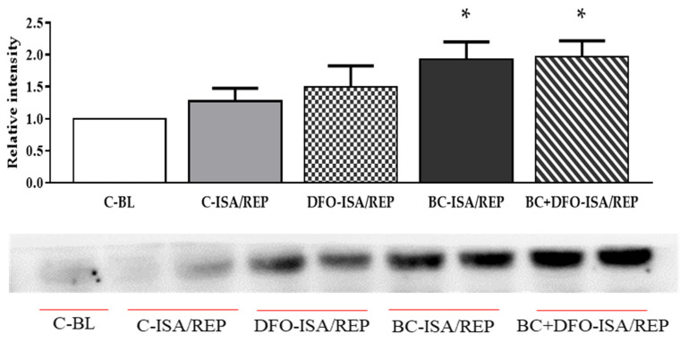
Figure 3.
Western blot analysis of HO-1 protein level in cardiac tissue. Expression of HO-1 protein in the hearts was measured in homogenized myocardial tissue samples from vehicle- or BC-treated rats with or without DFO treatment and ISA/REP injury. Tissue content of HO-1 is shown as a ratio of arbitrary units for HO-1 protein to total protein. Data are expressed as mean ± SEM of 7 different blots. * p < 0.05 compared to non-ischemic control (C-BL) hearts. A representative blot is also shown under the bars. Original blots are provided as supplementary Figure S1. C-BL: non-ischemic control group; C-ISA/REP: vehicle-treated, ischemic/reperfused control group; DFO-ISA/REP: vehicle-treated, ischemic/reperfused group with DFO administration; BC-ISA/REP: BC-treated, ischemic/reperfused group; BC + DFO-ISA/REP: BC-treated, ischemic/reperfused group with DFO administration.

