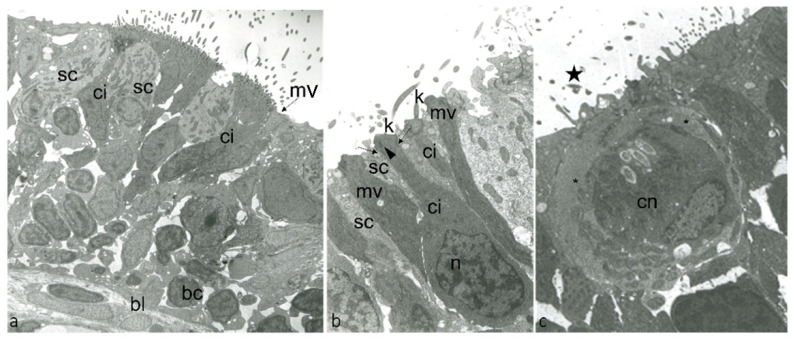
Figure 1.
Transmission electron micrographs of ultrathin sections showing the surface of olfactory epithelium adult zebrafish: (a) ci, ONR ciliated; mv, ONR microvillous; sc, supporting cells; bc, basal cells; bl, basal lamina. (b) ci, ONR ciliated; n,“checkerboard” nucleus of ciONR; K, kinocilia; mv, ONR microvillous; sc, supporting cells; kinocilium basal body foot (arrowhead), junctional complex with neighboring supporting cells (arrows). (c) cn, crypt neurons with several cilia within the crypt (star); special supporting cells (*).

