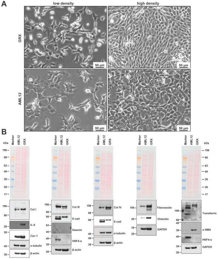
Figure 2.
Phenotypic characteristics of GRX cells. (A) GRX and AML12 cells were seeded in cell culture dishes and representative images taken from subconfluent and confluent cultures. Original magnifications are 200×. The space bars correspond to 50 µm. (B) Cell extracts were prepared from AML12 and GRX cells and analyzed for expression of α-smooth muscle actin (α-SMA), caveolin-1 (Cav-1), collagen type I (Col I), collagen type III (Col III), collagen type IV (Col IV), desmin, E-cadherin (E-cad), and fibronectin, hepatocyte nuclear factor 4 (HNF4-α), interleukin 6 (IL-6), transferrin, and vimentin. The expression of α-tubulin, β-actin, or GAPDH and the Ponceau S stain were included to demonstrate equal protein loading.

