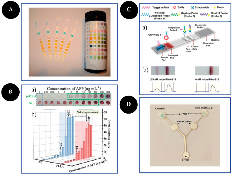
Figure 2.
Examples of different types of PADs. Dipsticks (A): urine test strips. Reproduced and adapted with permission from [13]; Spot test (B): photographs (a) and bar charts (b) of the corresponding grey values of the colorimetric readout based on a highly porous poly(L-lactic) acid nanofiber and NC platforms for AFP detection. Reproduced and adapted with permission from [30]; LFA (C): schematic representation of LFA principle for the detection of microRNA-215 (a) and photographs and corresponding optical measurements (b) of the developed LFA in the presence (2.5 nM) and absence (0 nM) of microRNA-215. Reproduced and adapted with permission from [32]; µPAD (D): photograph of the microfluidic sensor for detection of 1000 pM of microRNA-21 based on peroxidase mimetic activity of DNA-templated Ag/Pt nanoclusters. Reproduced and adapted with permission from [26].

