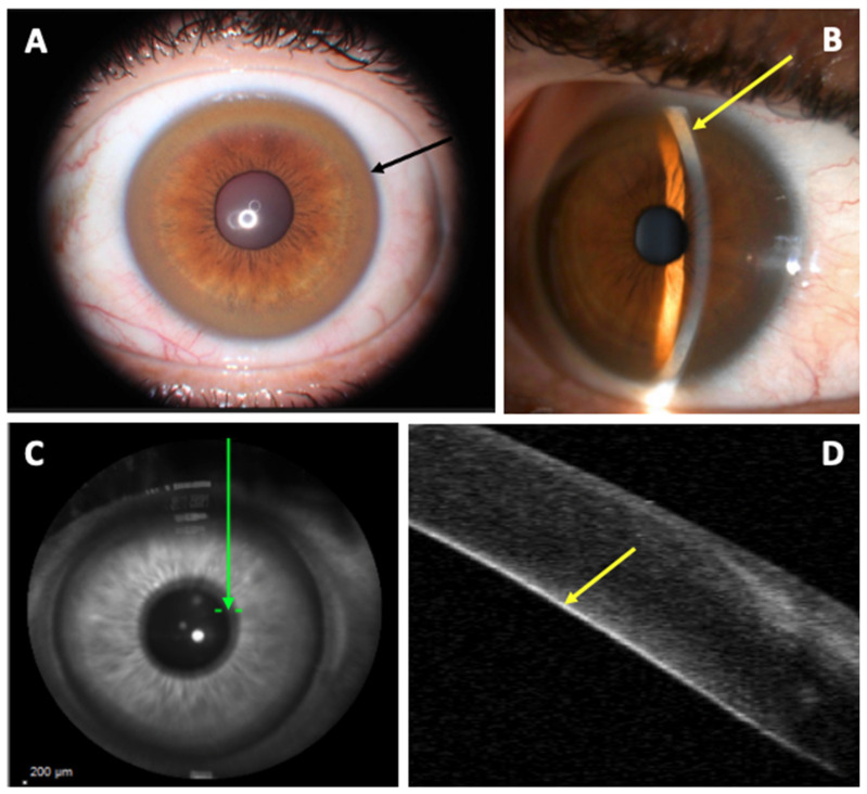
Figure 2.
Kayser–Fleischer Ring. (A): Slip-lamp examination showing a diffuse circumferential Kayser–Fleischer ring in the left eye (black arrow); (B): Slit-lamp examination: visualization of the copper deposit at the posterior part of the cornea in fine slit (yellow arrow); (C): Corneal B-scan localization (Spectralis; Heildelberg Engineering) (green arrow); (D): marked hyperreflectivity of the posterior part of the cornea corresponding to the copper deposit (yellow arrow).

