Fig. 1.
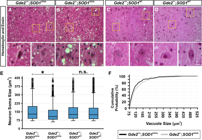
GDE2 is haploinsufficient in SOD1G93A transgenic animals. A-D Transverse sections of 14 week lumbar spinal cord ventral horns stained with H&E. Yellow boxes are magnified in lower panels, highlighting vacuolated and shrunken neuronal morphology in Gde2+/−;SOD1G93A mice. No morphological changes are seen in Gde2+/−;SOD1WT ventral horn neurons. Scale bar = 50 μm (A-D) or 15 μm (insets). E Box and whisker plot of neuronal soma sizes. Open circles are large diameter cells above the 95th percentile gates. Open triangles are > 3 times larger than the 75th percentile. Gde2+/−;SOD1G93A neurons are reduced in size compared to Gde2+/+;SOD1G93A controls, Kruskal–Wallis test (*p < 0.01). Gde2+/−;SOD1WT neurons are indistinguishable from Gde2+/+;SOD1WT controls, Kruskal–Wallis test (p = 0.588). No changes are detected in soma size between Gde2+/+;SOD1G93A and Gde2+/+;SOD1WT as expected at this timepoint [45]. F Cumulative probability distribution of vacuole sizes. Gde2+/−;SOD1G93A spinal cords contain larger vacuoles, Kolmogorov-Smirnoff test (p = 0.04). n = 4 Gde2+/+;SOD1G93A, 3 Gde2+/−;SOD1G93A, 3 Gde2+/+;SOD1WT, 4 Gde2+/−;SOD1WT

