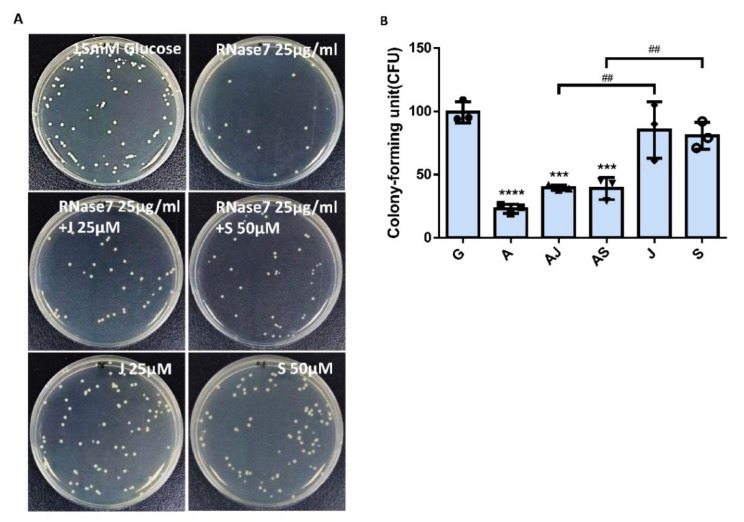
Figure 7.
JAK and STAT inhibitors affected the suppression of RNase 7 in UPEC-infected bladder cells in a high-glucose environment. SV-HUC-1 cells were pretreated with 25 μg/mL RNase 7 and simultaneously with 25 μM JAK inhibitor or 50 μM STAT inhibitor for 24 h, followed by UPEC infection (MOI:100) as described above. Cells pretreated with 25 μM JAK inhibitor or STAT3 inhibitor alone for 24 h followed by UPEC infection were used as respective controls. Cells infected with 15 mM glucose and UPEC alone were used as the respective positive controls. The definitions of each letter represented by the X axis are: C: control; G: with 15 mM glucose pretreatment alone; A: pretreated with 25 μg/mL RNase 7; AJ: coincubated with 25 μg/mL RNase 7 and 25 μM JAK inhibitor; AS: coincubated with 25 μg/mL RNase 7 and 50 μM STAT inhibitor; J: cells pretreated with 25 μM JAK inhibitor alone; S: Cells pretreated with STAT inhibitor alone. The picture shown is representative of a typical result. Twenty-four hours post-infection, all infected cells were (A) lysed and plated on LB agar and (B) measured using ImageJ software to analyze UPEC colonization. Colony-forming units (CFUs) were acquired after plating 10-fold dilutions of infected cells with lysis. Data are expressed as mean ± SD of three separate experiments. *** p < 0.001, **** p < 0.0001, compared to UPEC infection control groups. ## p < 0.01, compared between the two indicated groups.

