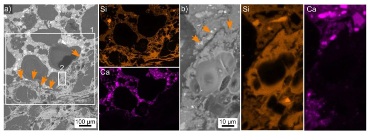
Figure 5.
(a) SEM micrograph featuring a cross section though the perlite-AAF composite presented in Figure 2b. The area in frame 1 was scanned by EDXS and element maps of Si and Ca are presented. Arrows highlight the boundary between perlite and the AAF. (b) The area in frame 2 presented in greater detail along with EDXS element maps of Si and Ca of this area.

