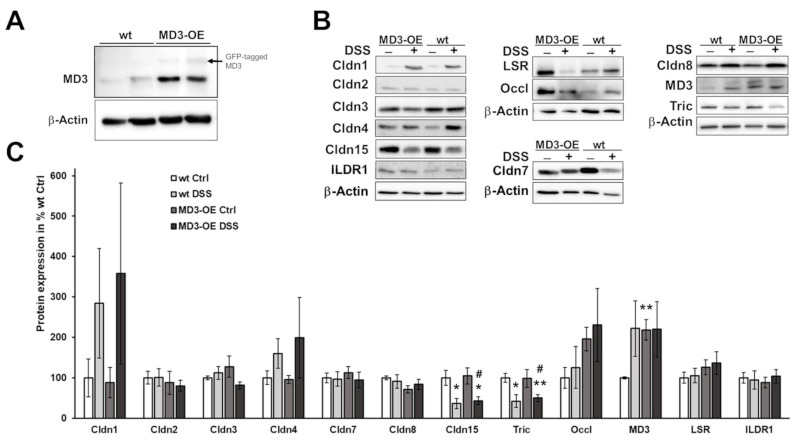
Figure 5.
Expression of MD3 and other TJ proteins. (A). Representative Western blots for MD3 and β-actin of wt mice and MD3-OE intestinal tissue. In addition to the MD3 signal in MD3-OE, some GFP-tagged MD3 was detectable. However, as the MD3 contained its start codon in addition to the N-terminal tag, amounts of untagged MD3 occurred in MD3-OE. (B). Representative Western blots for MD3 and other TJ proteins of wt and MD3-OE under control conditions and under the influence of DSS with corresponding β-actin signals. (C). Densitometric analysis of TJ expression profiles in wt and MD3-OE mice under control conditions (Ctrl) and DSS-induced colitis (n = 3–13; * p < 0.05; ** p < 0.01 compared to wt Ctrl; # p < 0.05 compared to MD3-OE Ctrl).

