
Fig. 1. Selective tau ablation in excitatory neurons reduces PTZ-induced epileptic activity in conditional knockout mice.
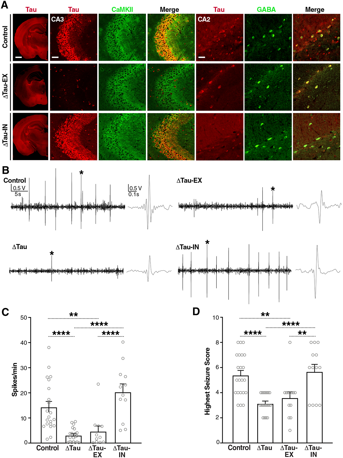
(A) Coronal brain sections from Maptflox/flox (Control), Emx1IRES-cre/+/Maptflox/flox (ΔTau-EX), and VgatIRES-cre/+/Maptflox/flox (ΔTau-IN) mice coimmunostained for tau (red) and markers (green) of excitatory (CaMKII) or inhibitory (GABA) neurons. The leftmost column shows hemibrains (scale bar, 900 μm) and the images on the right show details of the hippocampal CA3 and CA2 subfields (scale bars, 60 μm). CA3 illustrates tau ablation in excitatory neurons (middle row). Because CA2 contains fewer pyramidal cells, it is more suitable for illustrating tau ablation in inhibitory neurons (bottom row). Genotypes are indicated on the very left. (B to D) Video-EEG recordings were used to assess epileptiform spike activity (B and C) and behavioral seizure activity (D) in 3.5-month-old male Control, Mapt–/– (ΔTau), ΔTau-EX, and ΔTau-IN mice after intraperitoneal injection of PTZ (50 mg/kg). (B) Representative EEG traces recorded ~5 min after PTZ injection. Sections of traces marked with an asterisk are shown on the right at higher time resolution. (C) Epileptiform spikes per minute averaged from a 20-min EEG recording after the PTZ injection, or from shorter periods for mice that died during the recording. n = 11–21 mice per group. (D) Highest seizure score reached within 20 min after the PTZ injection. n = 13–22 mice per group. **P < 0.01 and ****P < 0.0001 by one-way ANOVA and Holm-Sidak test. Gray circles represent data from individual mice. Values in (C) and (D) are means ± SEM.