
Fig. 5 |. A stabilized actin cytoskeleton is required for myofibroblast persistence.
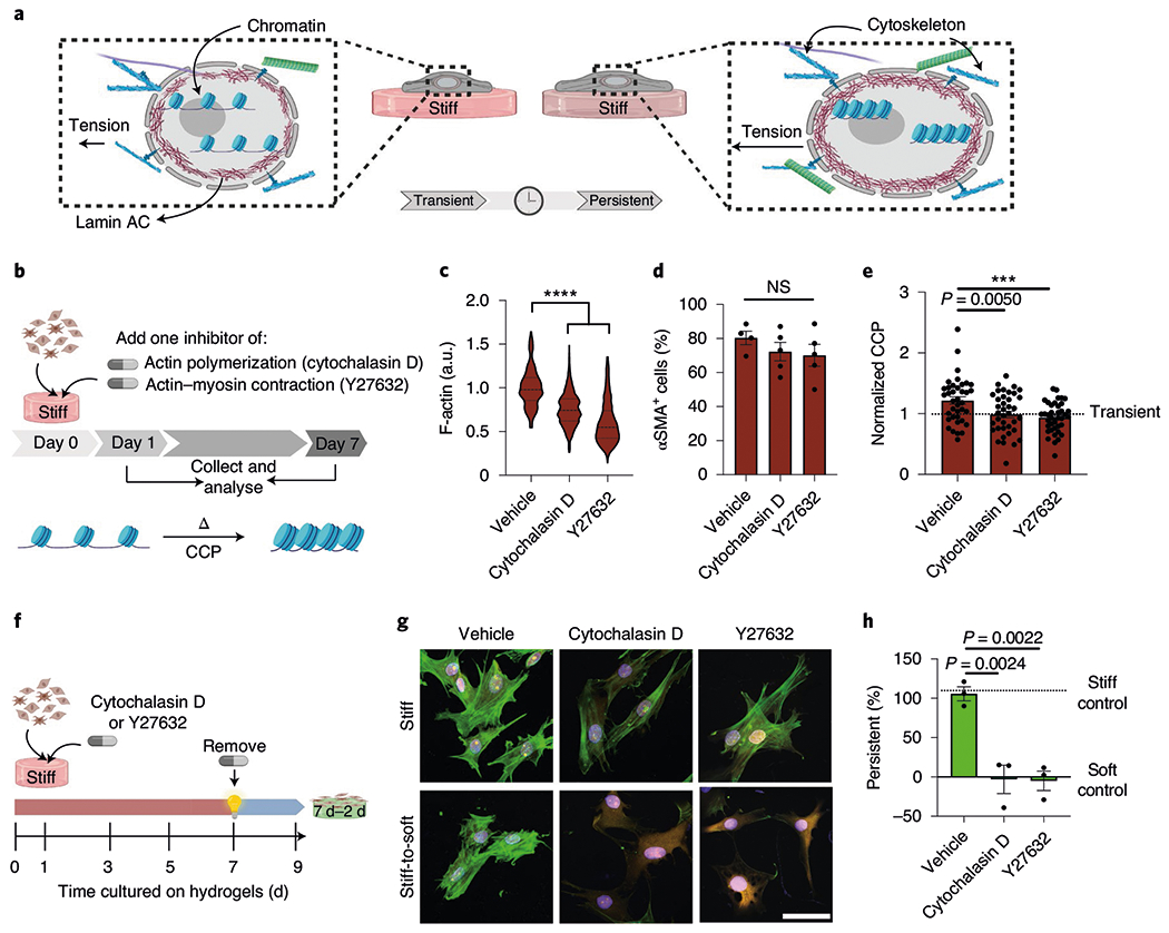
a, Illustration of the hypothesis that mechanics directly regulate chromatin structure in persistent myofibroblasts. b, Schematic of the cell culture experiment to test the effect of actin cytoskeleton on the chromatin structure. c, Actin levels determined from immunofluorescence in cells treated with the actin inhibitors cytochalasin D and Y27632 for 7 d on stiff hydrogels. The values were normalized to the vehicle. One-way ANOVA with Bonferroni’s post-hoc test; n = 192 (vehicle), 1,465 (cytochalasin D) and 1,300 (Y27632) cells; a.u., arbitrary units. d, Per cent αSMA+ cells after treatment with cytochalasin D and Y27632 for 7 d on stiff hydrogels. One-way ANOVA with Bonferroni’s post-hoc test; n = 4 (vehicle) and 5 (cytochalasin D and Y27632) hydrogels. e, CCP for persistent myofibroblasts (7 d stiff) treated with DMSO, cytochalasin D or Y27632 and normalized to CCP from transient myofibroblasts (1d stiff). One-way ANOVA with Bonferroni’s post-hoc test; n = 38 (vehicle) and 39 (cytochalasin D and Y27632) cells. f, Schematic of the cell culture experiment to test whether actin inhibition can prevent myofibroblast persistence. g, Representative images of persistent myofibroblasts treated with vehicle, cytochalasin D and Y27632 on stiff (9 d) or stiff-to-soft (7 d–2 d) hydrogels (n = 12 images per hydrogel). CellMask, yellow; αSMA, green; DAPI, blue. Scale bar, 50 μm. h, Per cent persistent myofibroblasts after treatment with vehicle, cytochalasin D and Y27632 (7 d stiff + drug, 2 d soft). One-way ANOVA with Bonferroni’s post-hoc test; n = 3 hydrogels. Data from three biologically independent replicates. ***P < 0.001; ****P < 0.0001; NS, not significant. Data reported as the mean ± s.e.m.