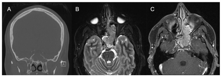
Figure 4.
IP-degenerated squamous cell carcinoma. (A) Non-contrasted CT sinus demonstrating a mass of the left sphenoid with erosion of the skull base at the sella. (B) STIR sequence contrasted MRI demonstrating an expansile mass originating from the left sphenoid lateral wall. (C) T1 sequence contrasted MRI with fat suppression demonstrating a mass of the left sphenoid and ethmoid cavity.

