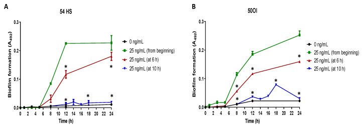
Figure 4.
Proteinase-3 addition at different stages of biofilm formation. Biofilm forming kinetics of a commensal (HS, healthy skin, panel (A)) and a clinical (OI, ocular infection, panel (B)) non-biofilm-forming isolate was done in the presence of proteinase-3 added at 6 or 10 h. The abundance of biofilm was determined according to Christensen et al. sampling at different points of the kinetics. Asterisk * indicates a significant difference (p < 0.05) concerning the bacteria with proteinase-3 since the beginning of the culture. The statistical analysis was performed using a one-way ANOVA with a Tukey’s test.

