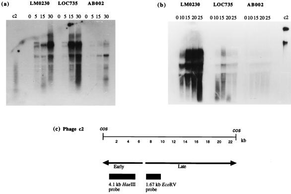
FIG. 5.
(a) Hybridization of total RNA isolated from phage c2-infected L. lactis subsp. cremoris LM0230, LOC735, or AB002 at 0, 5, 15, and 30 min postinfection with a 32P-labelled 4.1-kb HaeIII fragment from the early-expressed region. (b) Hybridization of total RNA isolated from phage c2-infected L. lactis subsp. cremoris LM0230, LOC735, or AB002 at 0, 10, 15, 20, and 25 min postinfection with a 32P-labelled 1.67-kb EcoRV fragment from the late-expressed region. In both panels, lanes labeled c2 contain phage c2 DNA used as a positive control. (c) Temporal transcription map of phage c2 with early- and late-expressed regions indicated; adapted from Beresford et al. (5).

