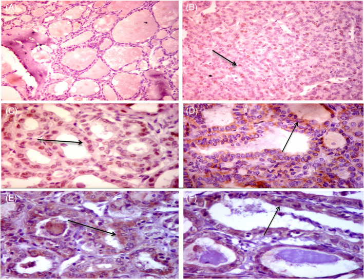
FIGURE 2.

Immunohistochemical (IHC) staining for p53 and BCL2 proteins in papillary thyroid carcinoma (PTC). IHC staining for p53 protein showed negative staining in non‐cancerous thyroid tissue (AX200) and scattered nuclear staining for p53 in PTC (Black arrow) (BX200). Diffuse nuclear staining for p53 in follicular variant of PTC (black arrow) (CX400). Staining for BCL2 protein showed focal mild cytoplasmic staining for BCL2 in hyperplastic nodule (DX 400), diffuse strong cytoplasmic staining for BCL2 in follicular variant of PTC (black arrow) (EX400), and moderate cytoplasmic staining in PTC (FX400)
