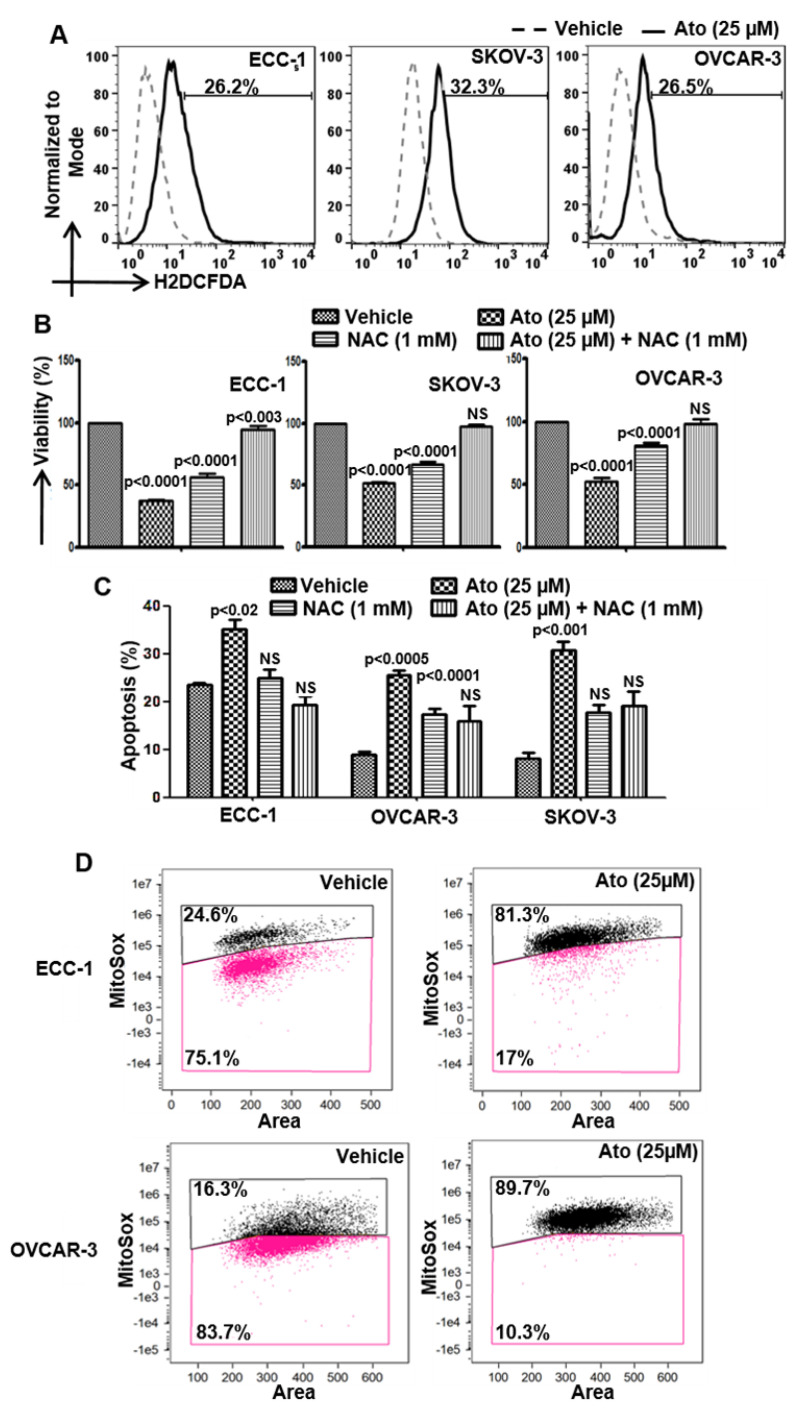
Figure 2.
Atovaquone induces oxidative stress in cancer cells. (A) Cells labeled with H2DCFDA were exposed to atovaquone (Ato) or DMSO (vehicle) and the intracellular oxygen radical flux was determined using flow cytometry. (B,C) Cells were preincubated with N-acetylcysteine (NAC) for 30 min prior to treatment with Ato and viability was monitored using the MTT assay and apoptosis by flow cytometry for Annexin V-FITC and propidium iodide. (D) Cells were pre-labeled with DRAQ5, Mitotracker green and Mitosox. After treatment with Ato or DMSO (vehicle), the radical flux was monitored by imaging cytometry. Data shown in all of the experiments (A–D) are representative of three independent experiments.

