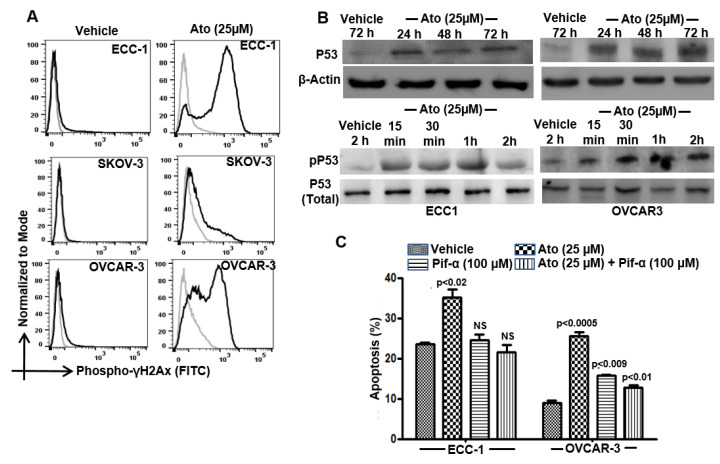
Figure 3.
Atovaquone activates p53. (A) Double-strand DNA breaks in the vehicle controls and atovaquone (Ato)-treated cells were determined by intracellular flow cytometry with FITC-conjugated anti-phospho-γ-H2Ax. (B) Phosphorylated (Serine-15 residue) and total p53 in control and atovaquone-treated cells were monitored by Western blotting. (C) ECC-1 and OVCAR-3 cells were treated with atovaquone in the presence/absence of the p53 inhibitor, pifithrin-α and monitored for proliferation (MTT assay) and viability and cell death (flow cytometry for Annexin V-FITC and propidium iodide) were monitored by flow cytometry. All data shown are representative of results from three independent repeats and statistical analysis compares atovaquone-treated cells with matching controls.

