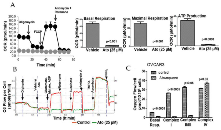
Figure 4.
(A). Inhibition of oxidative phosphorylation by Atovaquone (Ato) in OVCAR-3 cells was monitored on the Seahorse Xfe96 analyzer in triplicate. Data were normalized to the number of cells plated and cell viability was confirmed at the end of the experiment. Data for ECC-1 and SKOV-3 cells are shown in Supplemental Figure S4. (B). Complex-specific inhibition by atovaquone was monitored on an Oroboros analyzer. Digitonin was used to permeabilize the cells. Glutamate, malate and ADP enhance Complex I activity and succinate is a substrate for Complex II. TMPD and ascorbate are substrates for Complex IV. (C). The aggregate data from three independent experiments are shown to demonstrate that the addition of substrates for Complex I and II does not overcome the atovaquone-mediated inhibition of oxygen consumption rate (OCR).

