Figure 4.
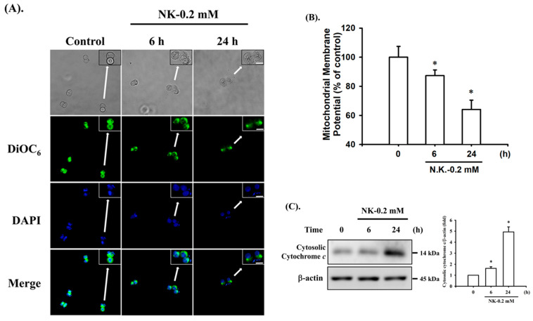
Norketamine (NK) induces mitochondrial dysfunction in RT4 cells. Cells were treated with or without NK (0.2 mM) for 6 and 24 h. (A) DiOC6 fluorescent probe was used to detect changes in MMP. The images were captured by fluorescence microscopy at a magnification of 400×. Scale bar: 20 μm. (B) Loss of MMP was detected and quantified using flow cytometry. Data are presented as the means ± SD of four independent experiments assayed in triplicate. (C) The release of cytochrome c from the mitochondria into the cytosolic fraction was analyzed using Western blot analysis. Results shown on a representative image, and quantification was determined by densitometric analysis. Each bar presented is the mean ± SD of three independent experiments. * p < 0.05 compared to vehicle control.

