Figure 4.
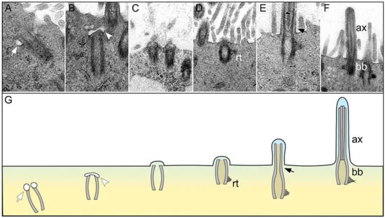
Ciliary regeneration in the trachea of a hamster 6 days after SARS-CoV-2 infection. (A–G) Representative transmission electron micrographs showing different stages of ciliary regeneration and a schematic illustration of the sequence of ciliary regeneration. (A) The centriole starts to produce a primary cilium by forming vesicles at the distal end (white arrows in (A,G)). (B) A ciliary bud develops at the distal end of the centriole invaginating the ciliary vesicle (white arrowheads in (B,G)). (C) The ciliary bud migrates to the apical pole of the cell and bulges the cell membrane. (D) The basal body develops a lateral rootlet (rt in (D,G)). (E) The organelle elongates apically and forms the axoneme (black arrows in (E,G)). (F) Mature cilia with regularly formed axoneme (ax in (F,G)) and basal body (bb in (F,G)). Magnifications are 31,500× in (A), 40,000× in (B), 10,000× in (C,D) and 12,500× in (E,F).

