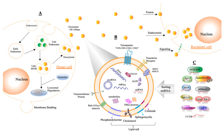
Figure 3.
Demonstrating the composition and generation of exosomes. (A) Exosomes are formed by endosomal multivesicular bodies (MVBs) budding inside the cell. Some of the MVBs that develop are delivered to the membrane of the cells after becoming late MVBs. MVBs can be dissolved during fusing with the lysosome or can release exosomes into the extracellular area by fusion with the plasma membrane through the exocytosis process, and the size range of exosomes is around ~50–150 nm. Acceptor cells receive exosomes through fusion, endocytosis, and/or signaling processes to insert their content. (B) Exosomes are enclosed by a phospholipid bilayer that contains a variety of components on its surface, such as tetraspanins (CD9, CD81, CD63, CD82), transferrin receptors, transmembrane proteins, molecular histocompatibility complex (MHCI, MHCII), Rab-GTPase annexin, and lipid rafts, while inward components contain biological species such as RNA (circRNA, mRNA, miRNA), proteins, DNA, and metabolites. In addition, tumor susceptibility gene 101 (TSG101) and apoptosis-linked gene 2-interacting protein X (ALIX) can be used as markers for exosomes. (C) Sorting miRNAs into exosomes can be regulated via different binding processes such as those of synaptotagmin-binding cytoplasmic RNA-interacting protein (SYNCRIP), sumoylated hnRNPA2B, Argonaute protein (Ago2), neutral sphingomyelinase 2 (nSMase2), major vault protein (MVP), CD63 with Y-box protein I (YBP1), Mex-3 RNA-binding family member C (MEX3C), protein 4A (Vps4A), lupus La protein (La protein), or the 3′ end of miRNA (3′UTR).

