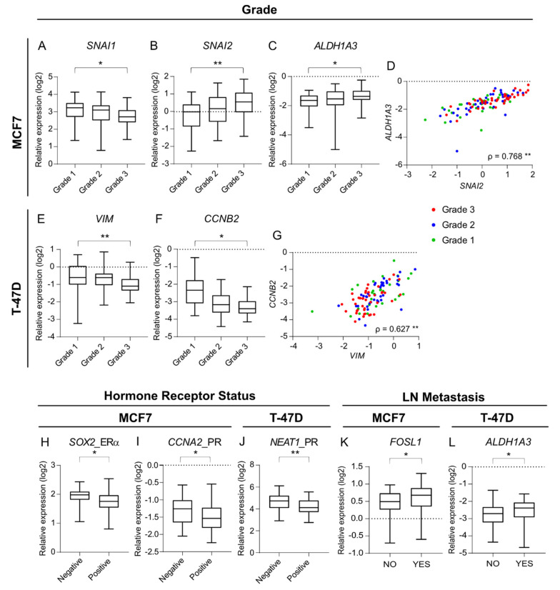
Figure 3.
Correlations between patient-derived scaffolds-induced expression of key markers in MCF7 and T-47D cells associate with cancer-related processes and clinico-pathological variables of the original tumor. (A–C,E,F) Boxplots (min to max) representing the median and the spread of the patient-derived scaffold (PDS)-induced expression values for the genes showing significant association to the clinical variable grade, in MCF7 (A–C) and T-47D cells (E,F). (D,G) Scatter plots illustrating the correlation between ALDH1A3 versus SNAI2 gene expressions in MCF7 for each PDS (D) and the correlation between CCNB2 versus VIM gene expressions in T-47D for each PDS (G), where colours denote the histological grade of the original tumor. (H–L) Boxplots (min to max) representing the median and the spread of the PDS-induced expression values for the genes showing significant association to the clinical variables progesterone receptor status (PR), estrogen receptor (ERα) status and presence of lymph node (LN) metastasis in MCF7 and T-47D cells (* p-value < 0.05, ** p-value < 0.01). Mann-Whitney U and Kruskall-Wallis statistical tests were performed for assessment of clinical variables (detailed information in Table 1). In the scatter plots, Spearman’s correlation coefficients (ρ) and the significance (** p-value < 0.01) are indicated (detailed information in Supplementary Figure S1 and Supplementary Table S5).

