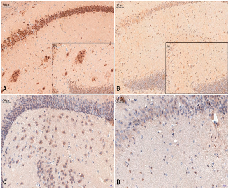
Figure 2.
Representative images of immunohistochemical reactions indicating β-amyloid (A,B) and TAU-5 (C,D) antigen expression were carried out on APP/PS1 + ovCYS group (A,C) and NCAR control (B,D) mouse brain. Nuclei are stained using hematoxylin. Magnification ×200 (A,B) and ×400 (C,D and insert).

