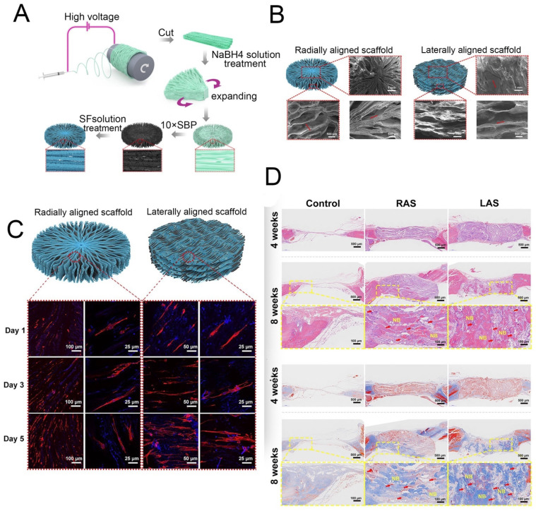
Figure 9.
(A) Schematic showing the process used to fabricate the 3D radially aligned nanofiber scaffolds. (B) Top and side scanning electron microscope images of the radially aligned scaffold. (C) BMSCs proliferated on the radially aligned scaffolds and linearly aligned scaffolds. (D) Histological analysis of the scaffolds after implantation for 4 and 8 weeks. Reprinted with permission from ref. [26]. copyright 2021 IPC Science and Technology.

