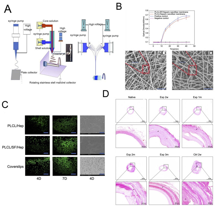
Figure 11.
(A) The schematic diagram of different electrospinning devices. (B) The ability to resist thrombosis. (C) Live cell staining photomicrograph of HUVECs and scanning electron microscope images of HUVECs grown on different materials at day 4. (D) The HE stains of different vascular scaffolds. Reprinted with permission from [128]. copyright 2019 Dove.

