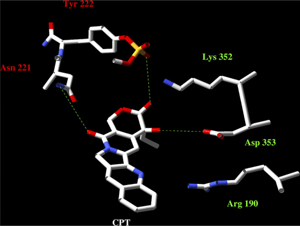
Fig. 7 –

Predicted 3D structure of the CPT binding pocket in LdTopIB. The homology modelling was made using the Swiss pdb database. The PDB ID 1t8i file corresponding to hTopIB − 70 kDa in a non-covalent complex with a 22-mer DNA duplex – was used as template. The final plots were obtained by using the MOLMOL programme. As the image displays, the residues of our study bound to the CPT E-ring (Asp-353) and the C17 (Asn-221); the presence of Arg-190 (Arg-364 in hTopIB) opens the possibility of interaction with the drug [32].
