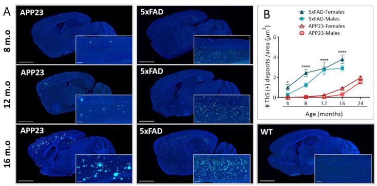
Figure 1.
Extracellular Aβ deposition in 5xFAD and APP23 mice. (A) Representative images of sagittal brain sections from female 5xFAD, APP23, and WT mice showing Aβ-positive fibrillar deposits stained with Thioflavin-S (ThS). Scale bar indicates 2 mm and 200 μm; m.o: months old. (B) Representation of the number of parenchymal Aβ deposits in male and female 5xFAD mice and APP23 mice at different ages. Data are expressed as the number of ThS-positive deposits (fibrillar Aβ deposits) per area. n = 4–7/group. Statistical differences were analyzed between APP23 and 5xFAD mice (females + males) and represented as: * p < 0.05, **** p < 0.0001.

