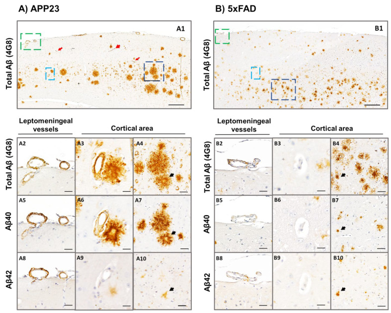
Figure 4.
Specific Aβ40 and Aβ42 immunodetection in the parenchyma and cerebral vasculature in APP23 and 5xFAD mice. Total Aβ (determined by anti-4G8), Aβ40, and Aβ42 immunodetection analyzed in brain sections of: (A) 16-month-old female APP23:and, (B) 5xFAD (mice. Zoom images from leptomeningeal vessels (green), cortical vessels (cyan), and amyloid plaques (dark blue) are shown in A2–A10 panels for the APP23 model and in B2-B10 panels for the 5xFAD model. A2–A4 and B2–B4 represent consecutive brain sections stained with anti-4G8 primary antibody; A5–A7 and B5–B7 represent consecutive brain sections stained with anti-Aβ40 primary antibody; and A8–A10 and B8–B10 represent consecutive brain sections stained with anti-Aβ42 primary antibody. Red arrows in A1 indicate amyloid-affected capillaries in APP23 brains. Black arrows in A4–A10 and B4–B10 indicate the same amyloid-plaques analyzed in consecutive sections using different primary antibodies. Scale bar in A1/B1 indicates 200 μm and in A2–A10 and B2–B10 indicates 40 μm.

