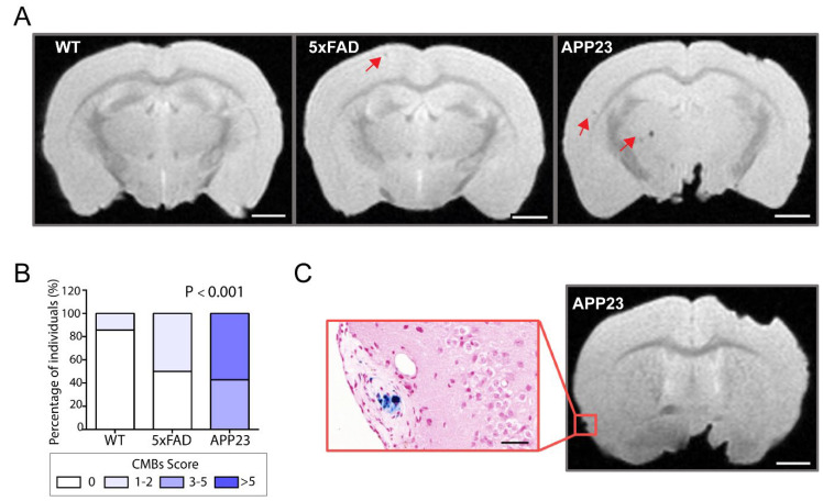
Figure 5.
MRI detection of CAA-related cerebral microbleeds in 5xFAD and APP23 mice. (A) Representative T2* magnetic resonance images (MRI) of cerebral microbleeds (cMBs) in 20-month-old male WT, 5xFAD, and APP23 mice. cMBs are indicated with red arrows. Scale bar indicates 2 mm. (B) Distribution of cMBs in WT, 5xFAD, and APP23 mice, according to the percentage of individuals affected. N = 4–7/group. (C) Comparison of cMBs in APP23 mice T2* sequences and Prussian blue staining showing iron hemosiderin deposits. Scale bar indicates 2 mm and 40 μm.

