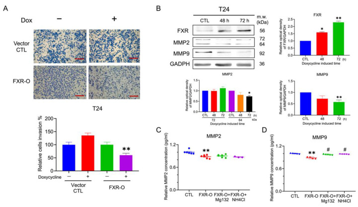
Figure 6.
The effect of invasive abilities after FXR overexpression. (A) Transwell invasion assays were performed for 16 h incubation in the T24 cells after FXR overexpression. The invasive cells were stained and captured. The right panel displayed the quantitative result. ** p < 0.01 compared to the control group. Scale bar = 200 μm. (B) The expression of matrix metalloproteinases-2 (MMP2) and matrix metalloproteinases-9 (MMP9) were analyzed by Western blotting in the T24 cells. GAPDH was used as the loading control. (C) The concentration levels of MMP2 in the CM of T24 cells were analyzed by ELISA. (D) The protein activity of MMP9 in the CM of T24 cells were analyzed by the Fluorokine assay. * p < 0.05; ** p < 0.01 compared with the control group. # p < 0.05 compared to the FXR-O group.

