Figure 3.
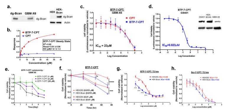
Cytotoxicity of BTP-7-CPT. (a) Western blot showing expression of dg-Bcan in patient-derived GBM-X6 cells, as well as in Bcan-overexpressing HEK cells (HEK-Bcan). (b) Binding kinetics analysis (steady-state) of BTP-7-CPT (red) and unmodified CPT (blue) to recombinant dg-Bcan protein using the ForteBio Octet system. Rmax (maximal response in response unit) and KD (dissociation constant) were calculated using a non-linear regression (one site specific binding) fit using the Graphpad Prism software. (c) Luminescent cell viability (CellTiter-Glo) of GBM-X6 cells treated with BTP-7-CPT (nwells = 3) for 72 h. (d) Luminescent cell viability (CellTiter-Glo) of GS401 recurrent GBM cells treated with BTP-7-CPT (nwells = 3) for 72 h. All IC50 values were measured through the non-linear ‘log(inhibitor) vs. response—variable slope (four parameters)’ fit. Western blot (right) shows higher expression of dg-Bcan in GS401 cells than in GBM-X6. (e) CellTiter-Glo assay of GBM-X6 cells treated with BTP-7-CPT at 1, 4, or 20 μM (nwells = 3) over 5 days. (f) CellTiter-Glo assay of HEK293 (red) or Bcan-overexpressing HEK (HEK-Bcan) cells (blue) treated with BTP-7-CPT at 0.015, or 41 μM (nwells = 3) over 3 days. (g,h) CellTiter-Glo assay of HEK-Bcan cells (blue) and control HEK293 cells (red) in the presence of (g) BTP-7-CPT or (h) Scr-7-CPT after 72 h. Statistical significance was calculated using a two-way ANOVA, Sidak’s multiple comparisons test (* p < 0.05, *** p < 0.001, **** p < 0.0001).

