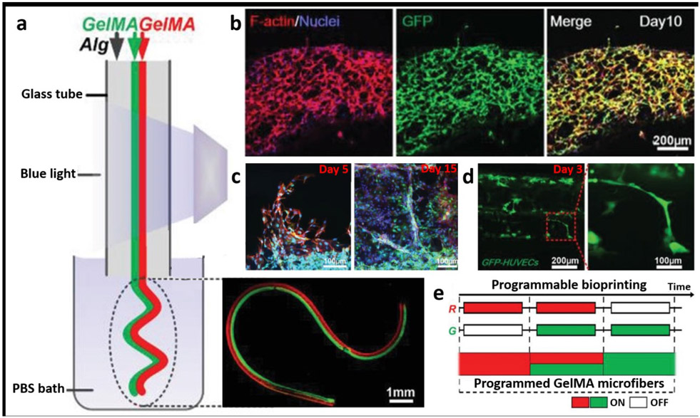
Figure 3:
a) Schematic depiction of the fabrication process for heterogeneous GelMA microfibers (Janus structures), b) 10 day culture of GFP+HUVEC-laden GelMA microfibers showing f-actin/nuclei staining in the constructs, c) Visualization of angiogenic sprouting in cell-laden constructs at different time points – it was noted that HUVEC-laden microfibers and MDA-MB-231-laden microfiber encapsulated in GelMA showed that there was an increase in sprout length with successive co–cultures, d) Visualization of vascular organoids and sprouting, e) a program layout for bioprinting of GelMA microfibers. Adapted with permission from reference [63].

