Figure 4.
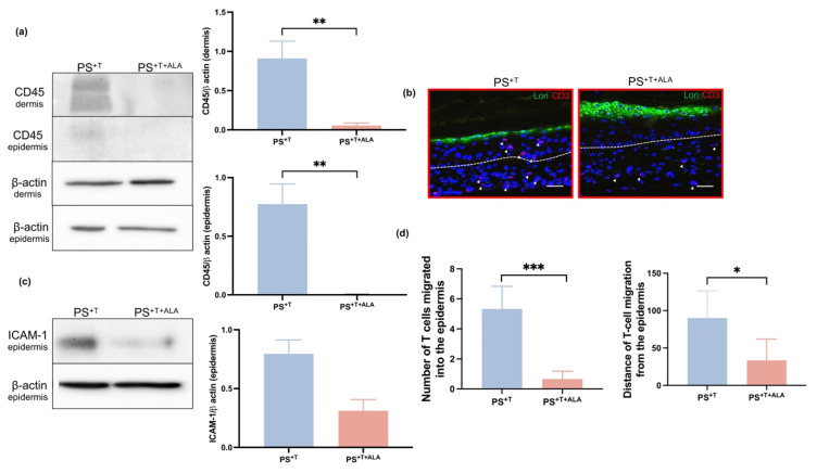
Expression of T cell markers in psoriatic skin substitutes. (a) Twenty micrograms of total protein from skin substitutes were analyzed by immunoblot for the presence of CD45 in the epidermis and the dermis of PS+T and PS+T+ALA. β-actin was used to control equal loading. One representative immunoblot is shown per protein (N = 3 donors per condition; n = 2 skin substitutes per donor); (b) Indirect immunofluorescence staining was conducted on PS+T and PS+T+ALA. The expression of CD3 is shown in red, while the expression of loricrin is shown in green and nuclei were counterstained with DAPI reagent (blue). The arrows show the CD3-labeled cells and the dashed white lines represent the basement membrane. Scale bars: 100 µm; (c) Twenty micrograms of total protein from skin substitutes were analyzed by immunoblot for the presence of ICAM-1 in the epidermis of PS+T and PS+T+ALA. β-actin was used to control equal loading. One representative immunoblot is shown per protein (N = 3 donors per condition; n = 2 skin substitutes per donor); (d) Number of T cells having migrated into the epidermis as well as their distance of migration in the epidermis (relative to the basal membrane) in PS+T and PS+T+ALA. Student’s t-tests were performed for statistical analyses. Significant differences are indicated by asterisks (* p < 0.05; ** p < 0.01; *** p < 0.001). Abbreviations: ALA: alpha-linolenic acid; ICAM-1: Intercellular adhesion molecule; Lori: Loricrin; T: T cells; PS+T: psoriatic substitutes produced with T cells; PS+T+ALA: psoriatic substitutes produced with T cells and supplemented with ALA.

