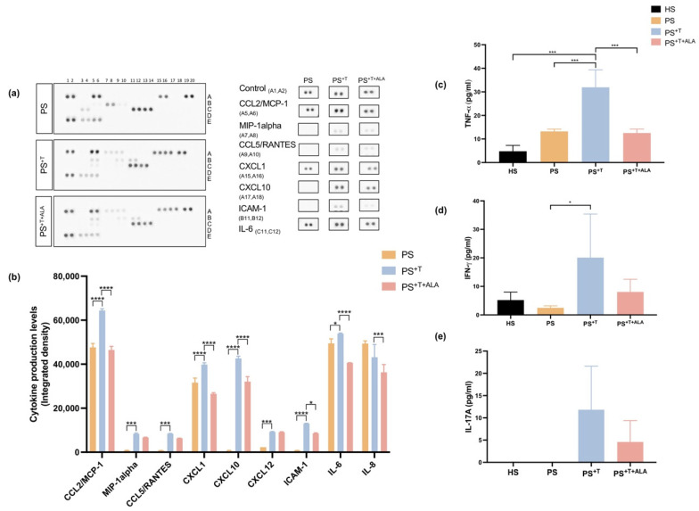
Figure 5.
Levels of inflammatory cytokines in PS, PS+T and PS+T+ALA. (a) Culture supernatants from PS, PS+T and PS+T+ALA were used to detect secreted cytokines using the Human Cytokine Array kit from R&D Systems. The duplicate spots correspond to the cytokines whose synthesis was the most altered following ALA supplementation; (b) Densitometric analysis of the dot blot duplicates from panel (a); (c) Tumor necrosis factor-α (TNF-α) levels in the culture medium of skin substitutes (N = 3 donors per condition, n = 2 culture supernatants per donor); (d) Interferon-gamma (IFN- γ) levels in the culture medium of skin substitutes (N = 3 donors per condition, n = 2 culture supernatants per donor); (e) IL-17A levels in the culture medium of skin substitutes (N = 2 donors per condition, n = 1 culture supernatant per donor). Statistical significance was determined using two-way ANOVA followed by Tukey’s post hoc test. Significant differences are indicated by asterisks (* p < 0.05, *** p < 0.001, **** p < 0.0001). Abbreviations: ALA: alpha-linolenic acid; CCL: chemokine ligand; CXCL: chemokine C-X-C motif ligand; HS: healthy substitutes; ICAM: intercellular adhesion molecule; IL: interleukin; T: T cells; PS: psoriatic substitutes; PS+T: psoriatic substitutes produced with T cells; PS+T+ALA: psoriatic substitutes produced with T cells and supplemented with ALA.

