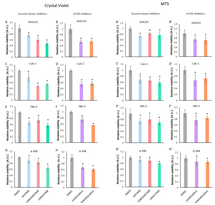
Figure 1.
Proliferation of HEK293, Caki-1, 786-O, and A-498 cells upon treatment with tyrosine kinase inhibitors or with mTOR inhibitors. Crystal violet (CV) staining (A–H) and MTS assays (A’–H’) were used to measure viability of cells upon treatment with tyrosine kinase and mTOR inhibitors after 72 h. Concentrations used were 1 μΜ Axitinib, 1 μΜ Cabozantinib, and 1 μΜ Lenvatinib in HEK293 cells (A,A’); and 20 μΜ Axitinib, 8 μΜ Cabozantinib, and 20 μΜ Lenvatinib in Caki-1 (C,C’), 786-O (E,E’), and A-498 (G,G’) cells. Everolimus and Temsirolimus were used at 0.1 μΜ in all cells (B,B’,D,D’,F,F’,H,H’). Note that HEK293 cells were more sensitive to the TKI treatments than the other renal cancer cells. Data are shown as relative proliferation ± S.D. Statistically significant results (p < 0.05) are marked with *. All data were normalized relative to untreated cells and are shown in arbitrary units (A.U.).

