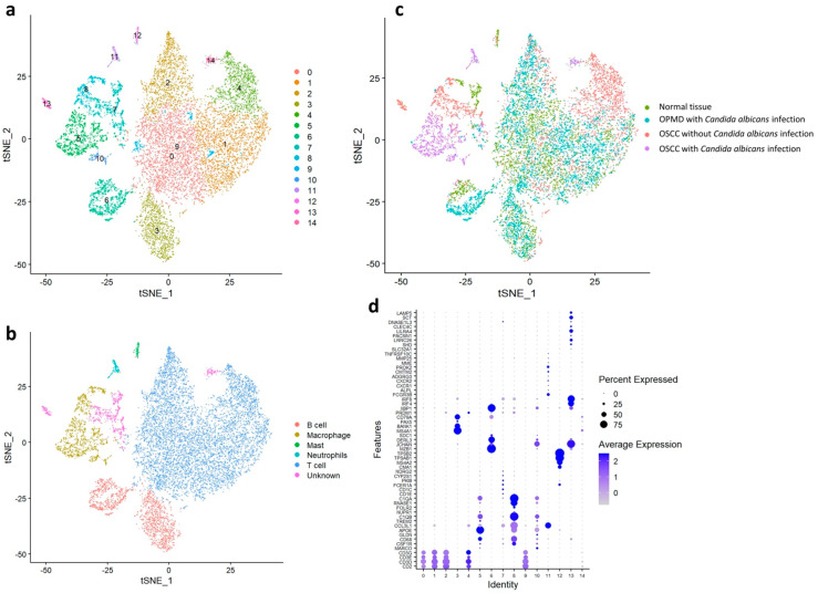
Figure 3.
Immune cell distribution in the normal tissue, OPMD lesion with Candida albicans infection, OSCC tissue with C. albicans infection, and OSCC tissue without C. albicans infection. (a) Classification of immune cells into 15 subtypes by using the Louvain algorithm. (b) Classification of immune cells into B cells, macrophages, mast cells, neutrophils, T cells, and unknown cells. (c) Separation of the four major groups. (d) Classification of cell types in each cluster by using specific biomarkers.

