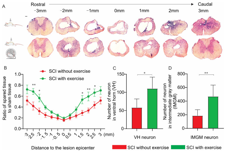
Figure 6.
Locomotor exercise spares spinal-cord tissue. Morphometric analysis of spinal-cord lesion. (A) HE staining of cross-sections of injured spinal cord at various distances from injury epicenter (both rostrally and caudally) for all experimental groups at 16th week postinjury. (B) Percentage of spared tissue calculated by normalizing area of spared tissue to total cross-sectional area of sham spinal cord. In the lesion epicenter, there was no difference in the spared tissue between groups with and without exercise. In the location rostral and caudal to the lesion center, exercise group had more spared spinal tissue. (C,D) Group with exercise had more neurons in the ventral horn (VH) and in intermediate gray matter (IMGM) than the group without exercise did. *: p < 0.05; **: p < 0.01; Scale bar 100 µm.

