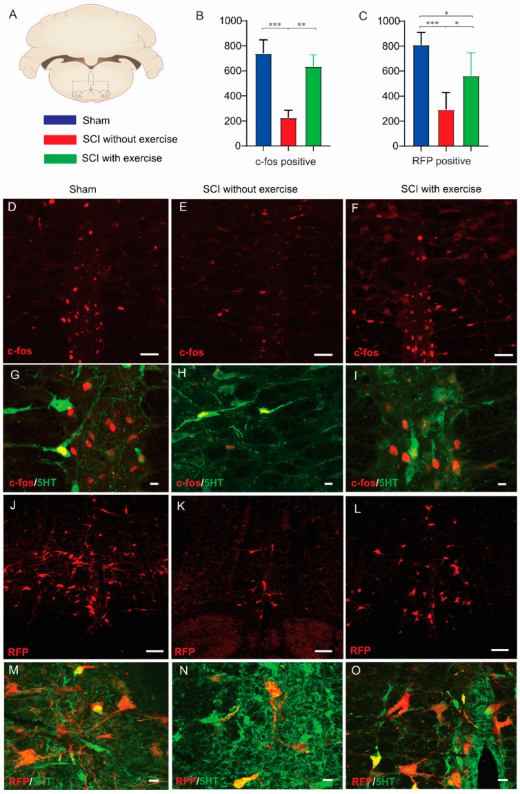
Figure 8.
Locomotor exercise enhances supraspinal-EUS motoneuron neural circuit. (A) Coronal section of the brain with locations of raphe nuclei (red box area) where imaging and quantification were performed. (B,C) Number of c-fos positive cells and red fluorescence protein (RFP)-labeled neurons in SCI without exercise group were significantly less than those in the sham and exercise groups. These results indicate that exercise training recruited more supraspinal neurons to control EUS function evidenced by more activated brain stem neurons during urodynamic experiment. (D–F) Immunofluorescence staining showing c-fos positive cells in coronal section of the brain. (G–I) Some (green arrow) but not all c-fos positive neurons (red arrow) are serotoninergic (c-fos+, red; 5-HT+, green). Subgroup of rats was injected with pseudorabies virus expressing RFP into the EUS. At 72h after injection, photomicrographs from coronal section of the brain demonstrate that RFP-labeled neurons were detected in the brainstem (J–L). (M–O) Some (green arrow) but not all of the RFP-labeled neurons (red arrow) are serotoninergic (RFP+, red; 5-HT+, green). Result of negative control from primary antibody omission presented as a Supplementary Figure S1. Scale bars are 100 µm in (D,E,F,J,K,L); 20 µm in (G,H,I,M,N,O). *: p < 0.05; **: p < 0.01; ***: p < 0.001.

