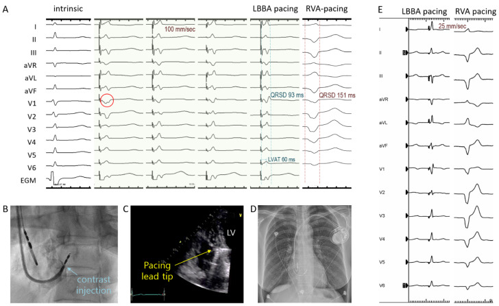
Figure 2.
Twelve-lead surface electrocardiogram and intracardiac electrogram (EGM) during intrinsic rhythm, unipolar LBBA pacing, and RVA pacing (A). Ideal location of LBBA shows a wide “W” shape QRS complex in V1 lead (red circle). Contrast injection (B) and echocardiography (C) showing the pacing lead tip implanted into the interventricular septum. After implantation of the LBBA pacing system (D), much narrower QRS duration was achieved by LBBA pacing (Left), compared with RVA pacing (Right) in the same patient (E). LBBA, left bundle branch area; LVAT, left ventricular activation time; QRSD, QRS duration; RVA, right ventricular apex; LV, left ventricle.

