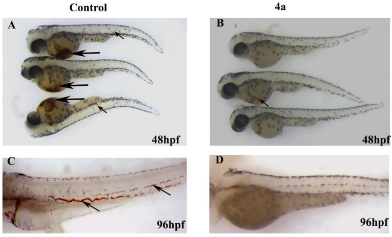
Figure 9.
Representative images of zebrafish embryos showing the O-dianisidien staining of erythrocytes. (A) Group of three control embryos at 48 hpf showing strong dianisidien-positive cells in the duct of Cuvier, indicated by black arrows, and also in the dorsal aorta and cardinal vein (trunk region by arrowhead). (B) Embryos treated with compound 4a (15.88 µM) did not stain positive for O-dianisidien (top and bottom embryos) or stained very weakly (middle embryo). (C) Mock-treated zebrafish embryos stained with O-dianisidien at 96 hpf marked all the erythrocytes (black arrows). (D) No positive O-dianisidien staining was detected in zebrafish embryos treated with compound 4a at 96 hpf.

