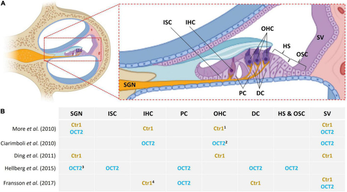
FIGURE 1.
Location of Ctr1 and OCT2 in inner ear cells. (A) Midmodiolar cross section of the cochlea with emphasis on the organ of Corti. Different cell types are indicated as follows: SGN, spiral ganglion neurons; ISC, inner sulcus cells; IHC, inner hair cells; PC, pillar cells; OHC, outer hair cells; DC, Deiters’ cells; HS, Hensen’s cells; OSC, outer sulcus cells; SV, stria vascularis. (B) Variable and inconsistent localization of Ctr1 and OCT2 in the inner ear in various studies; 1 base of OHC, 2 apical pole of OHC, 3 SGN type 1, 4 IHC synapse area.

