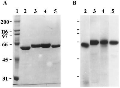
FIG. 1.
SDS-PAGE and autoradiography analyses of purified Cry1 toxins. (A) Coomassie blue-stained SDS-PAGE gel. Lane 1, molecular size markers; lane 2, Cry1Ac; lane 3, Cry1Ca; lane 4, Cry1Fa; lane 5, Cry1Ba. (B) Autoradiograph of 125I-labeled toxins. Lane 2, Cry1Ac; lane 3, Cry1Ca; lane 4, Cry1Fa; lane 5, Cry1Ba. The numbers on the left are molecular masses (in kilodaltons).

