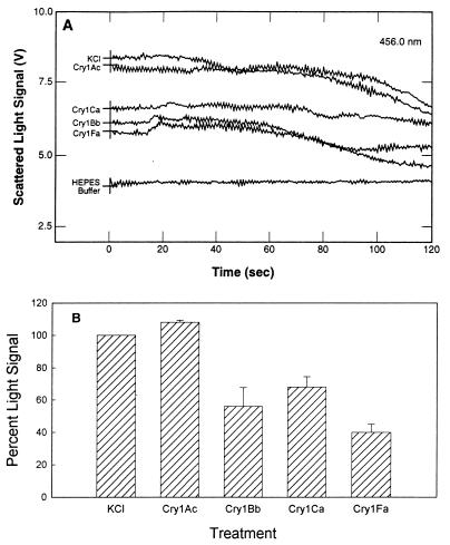
FIG. 5.
Scattered light signal of toxin-treated S. frugiperda BBMV after an increase in medium osmolarity. (A) Direct traces of scattered light from toxin-treated BBMV. BBMV were resuspended at a concentration of 0.5 mg/ml in 10 mM HEPES–Tris (pH 8.0) containing 0.1% BSA. Aliquots (1 ml) of BBMV were incubated with toxins (2.5 μg/ml) at room temperature for 30 min. Assays were initiated by simultaneously injecting 35 μl of a toxin-BBMV mixture and 35 μl of 0.5 M KCl into a cuvette in the spectrofluorimeter sample compartment. Light scattered at 90° from incidence was monitored for 120 s by using five measurements per s. In the HEPES sample, untreated BBMV were coinjected with 10 mM HEPES–Tris (pH 8.0) containing 0.1% BSA. The KCl sample contained untreated BBMV coinjected with 0.5 M KCl. (B) Proportion of scattered light signal change for toxin-treated S. frugiperda BBMV compared to KCl-treated BBMV, calculated from three independent assays. Five scattered light signal values (in volts) were extracted from raw data obtained at 1, 10, 20, 30, and 40 s in each assay, and then an average was calculated from these values. The standard deviations were calculated from the means of the three independent assays and are indicated as error bars.

